Improving Clinical Outcomes of Lumbar Fusion for Osteoporotic Patients
/By Michael Blatt
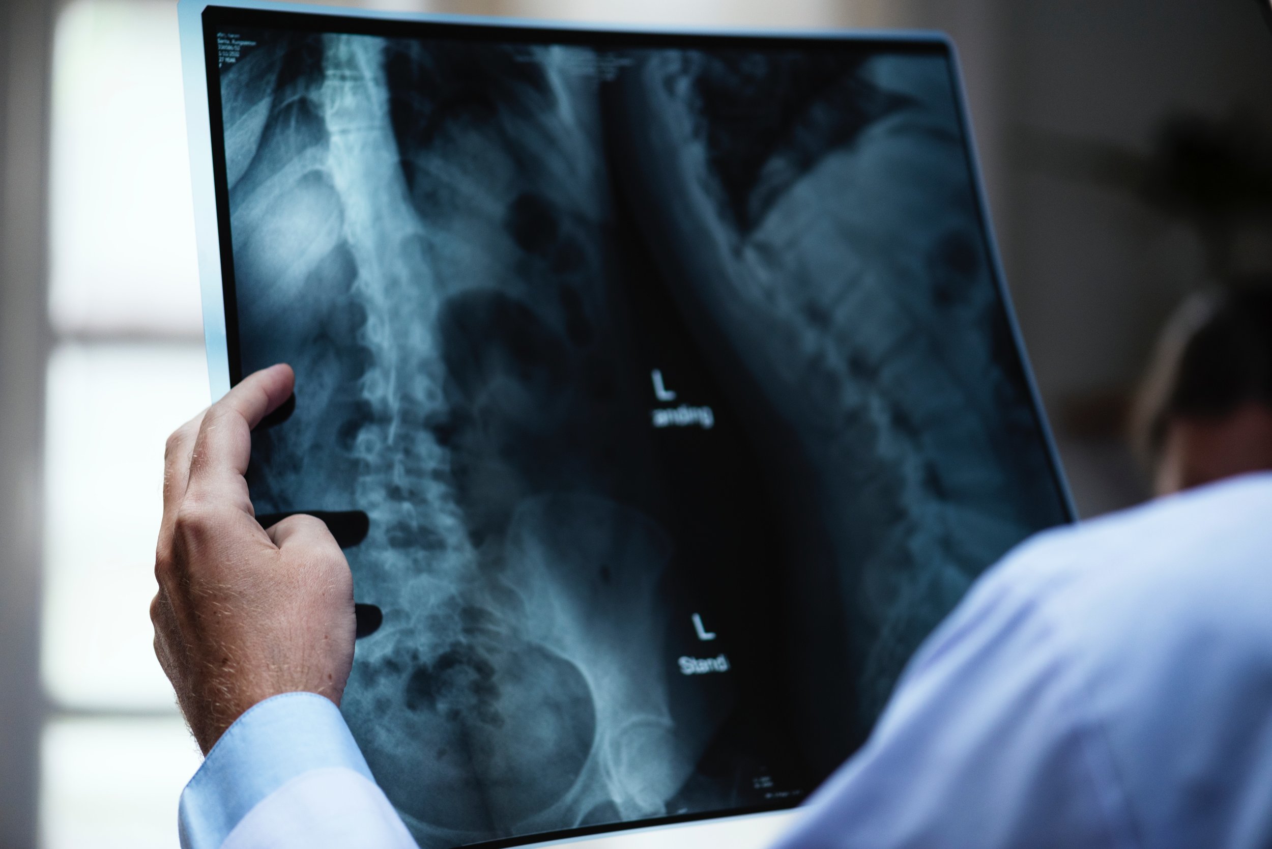
Picture yourself building a skyscraper in downtown Los Angeles with nothing but titanium-reinforced I-beams and concrete that is so brittle, even the slightest touch could make the foundation shatter. This is analogous to what spine surgeons are facing when patients with osteoporosis are in need of spinal fusion and/or fixation from trauma, degeneration, or spinal stenosis. Given the general surgical risk of spinal decompression and fusion compounded by the inability of the osteoporotic spine to act as anchor points for the spinal fusion, surgeons are in need of various multidisciplinary approaches in order to improve lumbar fusion outcome in this population subset. The purpose of this paper is to highlight the concerns that physicians face in assessing risk of patients with osteoporosis and the various therapeutic approaches for improving clinical outcome of spinal fusion.
Osteoporosis is a phenomenon that occurs when the body’s ability to grow new bone occurs slower than the resorption of old bone material—conventionally referred to as negative bone remodeling. In 2007, it was reported that in patients over 50, the rate of occurrence for osteoporosis in males and females was 14.5% and 51.3% respectively (Chin et al., 2007). As of 2014, it is estimated that approximately 54% of the US population older than 50 years old have osteoporosis or lowered bone mass, and it is predicted that the incidence of osteoporosis will increase as the population ages (Wright et al., 2014). This significant difference in male and female populations is due to the adverse effects associated with the loss of estrogen in menopause, which results in the loss of many cellular constituents involved in osteoclast development (Manolagas, 2000). It has been reported that patients being treated for spondylolisthesis have a significantly lower bone mineral density than patients being seen for spinal stenosis, and low-density bone may play a central role in the development of degenerative spondylolisthesis (Andersen et al., 2013). In patients who have undergone spinal fusion, osteoporosis-related bone disease is the primary cause of implant fixation failure, spinal fusion failure, and vertebral compression fractures adjacent to the site of fusion (Etebar & Cahill, 1999).
Spinal fixation is a procedure in which adjacent segments of the spine are mechanically bound together. At the lumbar levels of the spine, this is often performed posteriorly through the use of pedicle screws as anchor points for a continuous rod, which provides structural rigidity and local immobility for the levels of vertebrae fused. In the lumbar region of the spine, major structural deformities and associated pain can be relieved through fusion and fixation. The load that is typically placed on the lumbar vertebrae is then transferred from the center of the vertebral body to the axial rod and pedicle screws. However, if a patient’s spine has significantly low bone mineral density, perioperative measures must be taken to optimize the patient’s outcome.
Operatively, surgeons may consider using surgical implants that can passively promote osteointegration. The injection of polymethylmethacrylate (PMMA) through a cannulated pedicle screw has been demonstrated to be a safe and effective method of improving bone-union of pedicle screws for patients with severe osteoporosis (Moon, Cho, Choi, & Zhang, 2009). PMMA and other resins work by forming a biocompatible lattice around the interface of the pedicle screw and bone, which greatly improves the screw’s fixation in the bone. Chang, Liu, and Chen (2008) reviewed 291 cases of pedicle screws being augmented with PMMA, and found that no neurologic deterioration or bone cement leakage was present postoperatively in those cases. Hydroxyapatite coating of pedicle screws has also been shown to improve bone mineral density around the screw, providing exceptional fixation by reducing the risk of screw fixation with no significant difference in insertion torque (Yildirim, Aksakal, Hanyaloglu, Erdogan, & Okur, 2006).
Preoperatively, one strategy that is becoming more and more prominent is the usage of pharmacological approaches to limit bone resorption and induce bone remodeling. The goal of this approach is to chemically induce a decrease of bone resorption or locally increasing bone mineral density. Bisphosphonates are a class of drugs that have demonstrated an ability to induce apoptosis of osteoclasts, thus limiting the resorption of bone (Hughes et al., 1995). However, these drugs have also been shown to limit the union of grafted bone to the vertebral bodies, and thus appropriate postoperative treatment is required when bone grafts are used in surgery. (Takahata, Ito, Abe, Abumi, & Minami, 2008). There are also different pathways to target and inhibit various bone resorption pathways in postmenopausal patients (Roux, 2010). One of these resorption inhibitors, odanacatib (MK-0822), a human cathepsin K inhibitor, demonstrated increases in lumbar bone mineral density while being well-tolerated in postmenopausal women during phase I & II trials (Bone et al., 2010). The only known drug to act as an anabolic agent to osteoporosis is human parathyroid hormones (PTH), which at low and intermittent doses can increase bone formation while also inducing apoptotic effects on osteoclasts (Jilka et al., 1999). In a retrospective case analysis, administration of PTH1-34 postoperatively for a period of greater than six months was found to be effective in promoting bone union on posterolateral lumbar fusion (Ohtori et al., 2015).
Alternative to the usage of drugs that specifically inhibit the resorption of bone, there is a growing number of osteobiologic drugs and biomaterials that can actively promote bone growth (Hsu et al., 2017). Mesenchymal stem cells (MSCs), a class of pluripotent cells that are capable of differentiating into osteoblasts, can be derived from bone marrow aspiration or adipose tissue (Makino et al., 2018). It has been clinically demonstrated that embedding MSCs in a collagen scaffold can promote spinal fusion augmentation and improve clinical outcomes (Yousef, La Maida, & Misaggi , 2017). Bone morphogenic proteins (BMPs) have been demonstrated to play a significant role in the differentiation and proliferation of mesenchymal stem cells (Kannan, Dodwad, & Hsu, 2015). In a meta-analysis by Simmonds et al. (2013), recombinant human bone morphogenic protein-2 (rhBMP-2) was demonstrated to increase fusion rates and reduce pain. However, the increased risk of cancer associated with its usage remains of great significance to researchers (Carragee et al., 2013). In addressing this, researchers have found that the combined administration of PTH with BMP can improve fusion success rates while mitigating cancer risks associated with BMPs in rat models (Kaito et al., 2016). Two particular antibodies, anti-sclerostin antibodies and anti-RANKL monoclonal antibodies, are considered promising in the stimulation of bone growth; however, further studies are needed to determine their safety and effectiveness in spinal fusion (Makino et al., 2018).
For over 30 years, researchers have investigated the use of noninvasive electrical stimulation for the enhancement of spinal fusions (Gan & Glazer, 2006). By utilizing the piezoelectric properties of collagen, the induced mechanical strain can be used to stimulate bone growth as described by Wolff’s law. In a review by the European Spine Society, electrical stimulation has been demonstrated in animal models and in-vitro to stimulate the expression of bone morphogenic proteins, growth factors, and increase the prevalence of cytosolic Ca 2+ (Gan & Glazer, 2006). In addition to electrical stimulation, induction of a magnetic field has also demonstrated an ability to promote arteriolar vasodilation (Smith, Wong-Gibbons, & Maultsby, 2004). In a double-blind prospective study by Risso-Neto et al. (2017), postoperative pulsed electromagnetic field (PEMF) stimulation was shown to be effective in a heterogenous group of patients at stimulating bone union. Apart from electrical stimulation, mechanical stimulation, such as low-intensity pulsed ultrasound therapy, has also been shown to increase the bone grafting fusion rates in animal models undergoing lumbar fusion (Wang, Li, & Chen, 2015).
Physicians must also consider that the presence of other medical conditions associated with osteoporosis may impact the patient’s ability to withstand anesthesia, surgery, and postoperative care. For example, current or past heavy smoking has been demonstrated to significantly reduce bone mineral density, while also causing pulmonary and cardiovascular disease (Ward & Klesges, 2001). This increases the risk of pneumonia, cardiac events, or stroke in the postoperative period. The surgeon should consider whether a consult with an internal medicine specialist is necessary in order to optimize the patient’s medical status prior to surgery. Additionally, surgical candidates can improve their outcome through weight-bearing activity (Shanb & Youssef, 2014).
While treatment methods for osteoporotic patients requiring spinal fusion is a growing field of interest for researchers, there are currently many approaches available to surgeons to obtain adequate clinical outcomes. Physicians can opt for multimodal solutions, which involve the employment of osteoporosis medications to increase bone mineral density and modified surgical hardware that enable better pedicle screw purchasing and union to bone. Identifying lifestyle choices that induce bone resorption, such as tobacco usage, should also be addressed with the patient in optimizing their care. There are currently several approaches that physicians have to mitigate the risks and surgical complications associated with spinal fusions of the lumbar spine for patients with osteoporosis; however, the research and development of innovative methods for inducing bone growth continue.
References
Andersen, T., Christensen, F. B., Langdahl, B. L., Ernst, C., Fruensgaard, S., Østergaard, J., ... & Helmig, P. (2013). Degenerative spondylolisthesis is associated with low spinal bone density: A comparative study between spinal stenosis and degenerative spondylolisthesis. BioMed research international, 2013. doi:10.1155/2013/123847.
Bone, H. G., McClung, M. R., Roux, C., Recker, R. R., Eisman, J. A., Verbruggen, N., ... & Ince, B. A. (2010). Odanacatib, a cathepsin‐K inhibitor for osteoporosis: A two‐year study in postmenopausal women with low bone density. Journal of Bone and Mineral Research, 25(5), 937-947.
Carragee, E. J., Chu, G., Rohatgi, R., Hurwitz, E. L., Weiner, B. K., Yoon, S. T., ... & Kopjar, B. (2013). Cancer risk after use of recombinant bone morphogenetic protein-2 for spinal arthrodesis. JBJS, 95(17), 1537-1545.
Chang, M. C., Liu, C. L., & Chen, T. H. (2008). Polymethylmethacrylate augmentation of pedicle screw for osteoporotic spinal surgery: a novel technique. Spine, 33(10), E317-E324.
Chin, D. K., Park, J. Y., Yoon, Y. S., Kuh, S. U., Jin, B. H., Kim, K. S., & Cho, Y. E. (2007). Prevalence of osteoporosis in patients requiring spine surgery: Incidence and significance of osteoporosis in spine disease. Osteoporosis international, 18(9), 1219-1224.
Etebar, S., & Cahill, D. W. (1999). Risk factors for adjacent-segment failure following lumbar fixation with rigid instrumentation for degenerative instability. Journal of Neurosurgery: Spine, 90(2), 163-169.
Gan, J. C., & Glazer, P. A. (2006). Electrical stimulation therapies for spinal fusions: Current concepts. European Spine Journal, 15(9), 1301-1311.
Hughes, D. E., Wright, K. R., Uy, H. L., Sasaki, A., Yoneda, T., Roodman, D. G., ... & Boyce, B. F. (1995). Bisphosphonates promote apoptosis in murine osteoclasts in vitro and in vivo. Journal of Bone and Mineral Research, 10(10), 1478-1487.
Hsu, W. K., Goldstein, C. L., Shamji, M. F., Cho, S. K., Arnold, P. M., Fehlings, M. G., & Mroz, T. E. (2017). Novel Osteobiologics and biomaterials in the treatment of spinal disorders. Neurosurgery, 80(3S), S100-S107.
Jilka, R. L., Weinstein, R. S., Bellido, T., Roberson, P., Parfitt, A. M., & Manolagas, S. C. (1999). Increased bone formation by prevention of osteoblast apoptosis with parathyroid hormone. The Journal of clinical investigation, 104(4), 439-446.
Kaito, T., Morimoto, T., Kanayama, S., Otsuru, S., Kashii, M., Makino, T., ... & Yoshikawa, H. (2016). Modeling and remodeling effects of intermittent administration of teriparatide (parathyroid hormone 1-34) on bone morphogenetic protein-induced bone in a rat spinal fusion model. Bone reports, 5, 173-180.
Kannan, A., Dodwad, S. N. M., & Hsu, W. K. (2015). Biologics in spine arthrodesis. Journal of Spinal Disorders and Techniques, 28(5), 163-170.
Makino, T., Tsukazaki, H., Ukon, Y., Tateiwa, D., Yoshikawa, H., & Kaito, T. (2018). The biological enhancement of spinal fusion for spinal degenerative disease. International journal of molecular sciences, 19(8), 2430.
Manolagas, S. C. (2000). Birth and death of bone cells: Basic regulatory mechanisms and implications for the pathogenesis and treatment of osteoporosis. Endocrine reviews, 21(2), 115-137.
Moon, B. J., Cho, B. Y., Choi, E. Y., & Zhang, H. Y. (2009). Polymethylmethacrylate-augmented screw fixation for stabilization of the osteoporotic spine: A three-year follow-up of 37 patients. Journal of Korean Neurosurgical Society, 46(4), 305.
Ohtori, S., Orita, S., Yamauchi, K., Eguchi, Y., Ochiai, N., Kuniyoshi, K., ... & Kubota, G. (2015). More than 6 months of teriparatide treatment was more effective for bone union than shorter treatment following lumbar posterolateral fusion surgery. Asian spine journal, 9(4), 573-580.
Risso-Neto, M. I., Zuiani, G. R., Cavali, P. T., Veiga, I. G., Pasqualini, W., Amato Filho, A. C. S., ... & Miranda, J. B. D. (2017). Effect of pulsed electromagnetic field on the consolidation of posterolateral arthrodeses in the lumbosacral spine: A prospective, double-blind, randomized study. Coluna/Columna, 16(3), 206-212.
Roux, S. (2010). New treatment targets in osteoporosis. Joint Bone Spine, 77(3), 222-228.
Shanb, A. A., & Youssef, E. F. (2014). The impact of adding weight-bearing exercise versus nonweight bearing programs to the medical treatment of elderly patients with osteoporosis. Journal of family & community medicine, 21(3), 176.
Simmonds, M. C., Brown, J. V., Heirs, M. K., Higgins, J. P., Mannion, R. J., Rodgers, M. A., & Stewart, L. A. (2013). Safety and effectiveness of recombinant human bone morphogenetic protein-2 for spinal fusion: a meta-analysis of individual-participant data. Annals of internal medicine, 158(12), 877-889.
Smith, T. L., Wong-Gibbons, D., & Maultsby, J. (2004). Microcirculatory effects of pulsed electromagnetic fields. Journal of orthopaedic research, 22(1), 80-84.
Takahata, M., Ito, M., Abe, Y., Abumi, K., & Minami, A. (2008). The effect of anti-resorptive therapies on bone graft healing in an ovariectomized rat spinal arthrodesis model. Bone, 43(6), 1057-1066.
Wang, J., Li, J. W., & Chen, L. (2015). Effect of low–intensity pulsed ultrasound on posterolateral lumbar fusion of rabbit. Asian Pacific journal of tropical medicine, 8(1), 68-72.
Wang, T., Dang, G., Guo, Z., & Yang, M. (2005). Evaluation of autologous bone marrow mesenchymal stem cell–calcium phosphate ceramic composite for lumbar fusion in rhesus monkey interbody fusion model. Tissue engineering, 11(7-8), 1159-1167.
Ward, K. D., & Klesges, R. C. (2001). A meta-analysis of the effects of cigarette smoking on bone mineral density. Calcified tissue international, 68(5), 259-270.
Hsu, W. K., Goldstein, C. L., Shamji, M. F., Cho, S. K., Arnold, P. M., Fehlings, M. G., & Mroz, T. E. (2017). Novel osteobiologics and biomaterials in the treatment of spinal disorders. Neurosurgery, 80(3S), S100-S107.
Wright, N. C., Looker, A. C., Saag, K. G., Curtis, J. R., Delzell, E. S., Randall, S., & Dawson‐Hughes, B. (2014). The recent prevalence of osteoporosis and low bone mass in the United States based on bone mineral density at the femoral neck or lumbar spine. Journal of Bone and Mineral Research, 29(11), 2520-2526.
Yildirim, O. S., Aksakal, B., Hanyaloglu, S. C., Erdogan, F., & Okur, A. (2006). Hydroxyapatite dip coated and uncoated titanium poly-axial pedicle screws: An in vivo bovine model. Spine, 31(8), E215-E220.
Yousef, M. A. A., La Maida, G. A., & Misaggi, B. (2017). Long-term radiological and clinical outcomes after using bone marrow mesenchymal stem cells concentrate obtained with selective retention cell technology in posterolateral spinal fusion. Spine, 42(24), 1871-1879.


